Let’s talk thermography!
Did you know that the human body has a VERY specific temperature pattern? And changes in this pattern, because of inflammation or disease, can be detected using thermal imaging? Thermography or Digital Infrared Thermal Imaging (DITI) is a non-invasive screening tool that uses a specialized camera to measure extremely subtle temperature changes on the skin’s surface that correlate physiologically to underlying dysfunction or pathology.
The temperature changes are based on how nerves stimulate the skin and can indicate different health issues. We are not looking for “hot spots” as most think but a subtle temperature gradient change from what is classified as “normal” or healthy. It’s quite mathematical and not just “eyeballing” color differences!
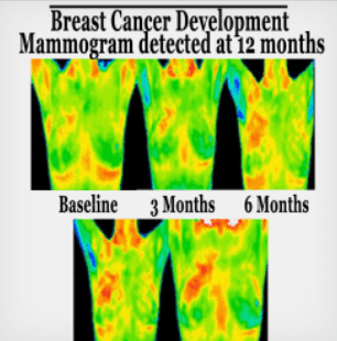
While other screening tools, such as mammography, MRI and ultrasound are looking for STRUCTURAL abnormalities (such as a tumor at least pea-sized), Thermography looks for FUNCTIONAL abnormalities (the process that occurs before a tumor grows) – such as new blood vessel development (“angiogenesis”). New blood vessels develop under VERY specific conditions – often when the body is trying to “grow something new”.
It’s best known for its role in breast health, but many are learning that it can be used to assess almost the entire body! Important to note – it’s not helpful directly for brain issues, however.
In addition, thermography can be helpful in differentiating the origin of pain anywhere in the body whether neurological, muscle, bone or cardiovascular providing more information for effective treatments.
There is no radiation, no skin contact and no discomfort. Thermography is useful in ANY condition. There are no contraindications and can be safely performed on anyone. For best results specifically for breast imaging it should be repeated 3 months after the initial scan to establish an individual baseline then once per year thereafter. We repeat initial breast imaging at 3 months to verify there are no changes in temperature gradients. In other words – vasculature of breasts should NOT change over time (unless a woman is pregnant and/or lactating!). We know that breast cancer cells significantly increase over a 3 month period and we want to look for those changes early rather than wait 6+ months – too long!!
While a client gets great information from their first thermography it excels when comparing multiple images over time. The initial set of images are then compared to the second set and the third set, etc. – over time this gives a better overall view of health – in essence it’s like “mapping” the body’s physiology each time.
Thermographic images are interpreted by Board-certified MD Clinical Thermologists. Much like a Radiologist, a Thermologist is trained to interpret images taken by a Thermographer using a thermal camera. These results are then presented in a report as interpretation of findings.
Some of the health issues we have found it helpful for:
BREAST CANCER

FIBROMYALGIA

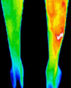
STRESS FRACTURE

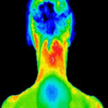
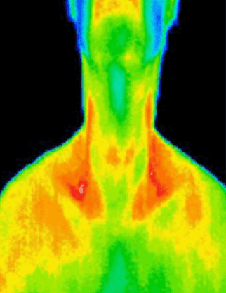
THYROID DISORDER

The FDA has made it mandatory for me to tell you that it shouldn’t be used as a stand-alone assessment – specifically for breast tissue.
Now I must admit – this is the ONLY thing I will agree with the FDA on! 😊
It is an adjunctive/supportive screening tool and is not a replacement for diagnostic tools (such as biopsies) but can provide crucial information on the origin of symptoms as well as identify pre-disease states resulting in early diagnosis. Research shows that thermal imaging can detect some cancers 8-10 years before they are visible on mammogram therefore allowing the golden opportunity for preventative treatments.
But for breast health – if someone feels a lump I ALWAYS recommend a breast ultrasound. Don’t rely on thermography (or mammography) only – EVER!
I have used thermography in my practice for 11 years now and ONLY used Meditherm equipment – it’s superior to any other system out there. The medical doctors who interpret our images are highly trained and represent a variety of medical specialties, including pathology, emergency medicine, OB/Gyn and more.
Like other inexpensive and effective things in natural medicine, Thermography is often demonized because it’s not a big money maker for the medical system like other screening tools, but it offers great information for those needing answers or wanting to have an annual assessment of health.
Downside is that it’s the only part of my practice that cannot be virtual ☹
I always recommend a Meditherm practitioner when searching for options closest to you! https://thermologyonline.org/
Big Hugs!!
Dr. K
Interested in chatting with us further? Call our office or click on the button below to schedule a zoom call!

